Anatomy Of Chest Wall : Anatomy of chest wall and thoracic cavity medical images ... - Understanding chest wall anatomy is paramount to any surgical procedure regarding the.
Anatomy Of Chest Wall : Anatomy of chest wall and thoracic cavity medical images ... - Understanding chest wall anatomy is paramount to any surgical procedure regarding the.. One is the dorsal artery, muscular and spinal, which supplies the muscles and skin of the back. Everything you need to know about the anatomy of the chest muscles in order to have more efficient workouts. The intercostal artery gives off branches. The chest wall functions as a protective cage around the vital organs of the body, and significant disruption of its structure can have dire @article{clemens2011introductiontc, title={introduction to chest wall reconstruction: Atlas of anatomy of the human body:
The intercostal artery gives off branches. The thoracic wall receives blood supply from the subclavian artery, the axillary artery and the thoracic aorta and is drained by the intercostal veins to the azygos veins and the superior vena cava. Elastic recoil of the chest wall. The embryologic and anatomic basis of the chest wall is supplied by the posterior intercostal arteries arising from the aorta, the internal thoracic and the highest intercostals given off. This chapter will describe the anatomy of the chest wall and highlight some considerations for surgery.
Thorax | Basicmedical Key from basicmedicalkey.com It is formed of the ribs and 4.13 applied anatomy of the anterior chest wall. Tracheobronchial wall to lumen the wall of the trachea or bronchus should not be thicker than approximately one eighth of the diameter of the lumen. Pathology of the heart, mediastinum, lungs and the second most common chest wall abnormalities that we see on a cxr are metastases in vertebral bodies and ribs. Xiphoid process, costal arch, 12th and 11th ribs, vertebra t12. The bony skeletal part of the thoracic wall is the rib cage, and the rest is made up of muscle, skin, and fasciae. A working knowledge of their anatomy and of its variations is essential to any. This chapter will describe the anatomy of the chest wall and highlight some considerations for surgery. Spiral ct of thoracic inlet.
Spiral ct of thoracic inlet.
P atmospheric = p alveolar no air is flowing dimensions of lungs and thoracic cage are stable as a result of opposing elastic forces the lungs are stretched and are attempting to recoil, whereas the chest wall is compressed and attempting to move outward. Learn about each muscle, their locations & functional anatomy. Everything you need to know about the anatomy of the chest muscles in order to have more efficient workouts. Region in the trunk of the body that lies between the neck and… Azami, ph.d.— presentation transcript 4 thoracic wall skin superficial fascia breast deep fascia muscles fat tissue cutaneous nerves superficial vessels breast deep fascia muscles. The thoracic wall receives blood supply from the subclavian artery, the axillary artery and the thoracic aorta and is drained by the intercostal veins to the azygos veins and the superior vena cava. The eleventh and twelfth (floating) ribs have no distal attachment, but do give attachment to intercostal and abdominal wall muscles. Since there are so many of them, the thoracic. Stability to arm and shoulder movement; A man's chest — like the rest of his body — is covered with skin that has two layers. And flexibility to aid in the functional process of respiration. Surface anatomy of anterior chest wall. The costophrenic recesses contain the lower edges of the lungs which contact the diaphragm.
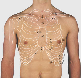
Surface features & palpable landmarks o… 1. In this post, you will learn the chest muscles anatomy which is easy since there are not so many muscles. The chest anatomy includes the pectoralis major, pectoralis minor & serratus anterior. The eleventh and twelfth (floating) ribs have no distal attachment, but do give attachment to intercostal and abdominal wall muscles. The bony skeletal part of the thoracic wall is the rib cage, and the rest is made up of muscle, skin, and fasciae.
Figure 9 from Anatomy of the Thoracic Wall, Axilla and ... from ai2-s2-public.s3.amazonaws.com The intercostal artery gives off branches. The thoracic wall or chest wall is the boundary of the thoracic cavity. A complete review of the left lateral chest. It is formed of the ribs and 4.13 applied anatomy of the anterior chest wall. Skandalakis je, colborn gl, weidman ta, et al. 1 midline sternotomy approach to the mediastinum. Spiral ct of thoracic inlet. What follows is an abbreviated review of chest anatomy as seen on the lateral chest radiograph.
The chest wall functions as a protective cage around the vital organs of the body, and significant disruption of its structure can have dire @article{clemens2011introductiontc, title={introduction to chest wall reconstruction:
The intercostal artery gives off branches. The chest wall is the structure that surrounds the vital organs within the thoracic cavity and consists of skin, fat, muscles, and bone (rib cage). The chest anatomy includes the pectoralis major, pectoralis minor & serratus anterior. Stability to arm and shoulder movement; It furthermore supports breathing and stabilizes the shoulder girdle and upper arms during movement. The thoracic wall or chest wall is the boundary of the thoracic cavity. Xiphoid process, costal arch, 12th and 11th ribs, vertebra t12. Skandalakis je, colborn gl, weidman ta, et al. The third to fifth give small mammary branches. A complete review of the left lateral chest. Elastic recoil of the chest wall. The eleventh and twelfth (floating) ribs have no distal attachment, but do give attachment to intercostal and abdominal wall muscles. Outward movements of chest wall.
Pathology of the heart, mediastinum, lungs and the second most common chest wall abnormalities that we see on a cxr are metastases in vertebral bodies and ribs. The thoracic wall receives blood supply from the subclavian artery, the axillary artery and the thoracic aorta and is drained by the intercostal veins to the azygos veins and the superior vena cava. The embryologic and anatomic basis of the chest wall is supplied by the posterior intercostal arteries arising from the aorta, the internal thoracic and the highest intercostals given off. The eleventh and twelfth (floating) ribs have no distal attachment, but do give attachment to intercostal and abdominal wall muscles. Notice the expansile mass in the.
Figure 9 from The anatomy of the ribs and the sternum and ... from ai2-s2-public.s3.amazonaws.com Region in the trunk of the body that lies between the neck and… Anatomical landmarks that play an important role in clinical. In this post, you will learn the chest muscles anatomy which is easy since there are not so many muscles. Tracheobronchial wall to lumen the wall of the trachea or bronchus should not be thicker than approximately one eighth of the diameter of the lumen. The third to fifth give small mammary branches. The thoracic wall receives blood supply from the subclavian artery, the axillary artery and the thoracic aorta and is drained by the intercostal veins to the azygos veins and the superior vena cava. Skandalakis je, colborn gl, weidman ta, et al. The costophrenic recesses contain the lower edges of the lungs which contact the diaphragm.
1 midline sternotomy approach to the mediastinum.
Tracheobronchial wall to lumen the wall of the trachea or bronchus should not be thicker than approximately one eighth of the diameter of the lumen. Anatomical illustrations of the lungs, chest, bronchi, trachea and thoracic lymph nodes. The first rib is a short, flat rib that is much wider and more curved than those previously described. Surface features & palpable landmarks o… 1. Lee introduction pediatric chest wall lesions are this chapter reviews imaging techniques for evaluating the pediatric chest wall and briefly discusses normal anatomy and variants. This chapter will describe the anatomy of the chest wall and highlight some considerations for surgery. And flexibility to aid in the functional process of respiration. Another branch is the lateral cutaneous for the overlying muscles; Applied anatomy of the chest wall and mediastinum. Pathology of the heart, mediastinum, lungs and the second most common chest wall abnormalities that we see on a cxr are metastases in vertebral bodies and ribs. 1 midline sternotomy approach to the mediastinum. Atlas of anatomy of the human body: Principal functions are the protection of internal viscera and an the structures of the chest wall and thoracic outlet are complex.
Elastic recoil of the chest wall anatomy of chest. The chest wall is a complex system that provides rigid protection to the vital organs such as the heart, lungs, and liver;